Research and Development
Background of cancer immunotherapy
While conventional anticancer treatments, in particular, chemotherapy, hormone therapy, targeted therapy and radiotherapy have been shown substantial benefit to selected groups of cancer patients, tumor relapse is a frequently encountered problem that results from tumor metastases and acquired resistance. Immunotherapy emerged as the next-generation anticancer therapy after decade-long efforts to understand and translate biological insight into lifesaving therapies. Cancer immunotherapy utilizes the body’s immune system to attack cancer. It can function basically in two clinical scenarios: 1) if the immune system does not recognize the tumor cells, a response must be induced to educate the recognition; 2) if the immune system can recognize but the response is not sufficient, it must be stimulated and/or enhanced.
There are three distinct approaches to cancer immunotherapy:
- monoclonal antibody-based therapies
- immune cell mediated therapies
- cancer vaccines

Immune checkpoint proteins
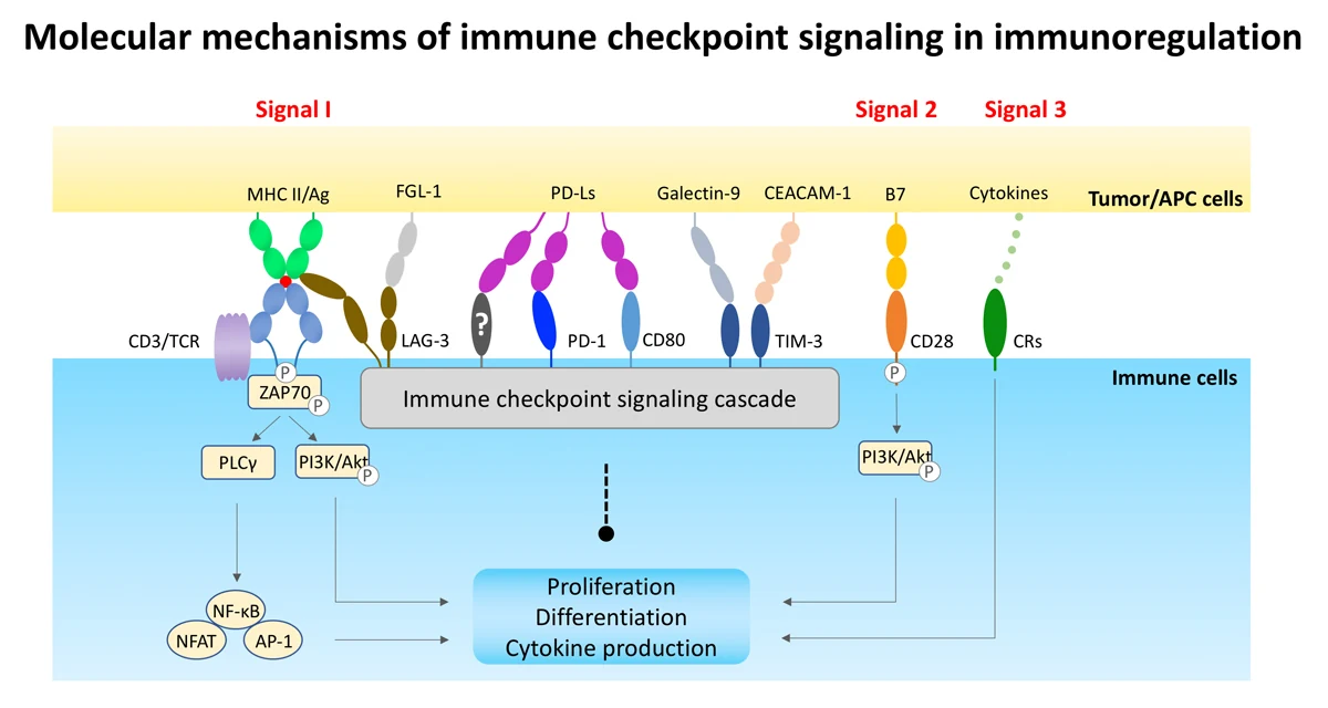
To avoid the collateral tissue damage from uncontrolled immune activation in peripheral tissues, the immune system has evolved to establish regulatory machinery to maintain self-tolerance and modulated duration and amplitude of immunity. This protective mechanism is mediated by a group of inhibitory signaling pathways better known as immune checkpoints. Fundamentally, immune checkpoint proteins act as inhibitory receptors on the immune cell surface and regulate cell fate through protein-protein interaction with their ligands on the surface of antigen-presenting cells and cancer cells. So far, a number of proteins on immune cell surface have been identified to play crucial roles in checkpoint signaling such as CTLA-4 (cytotoxic T-lymphocyte-associated Protein 4), PD-1 (Programmed cell death protein 1), TIM-3 (T-cell immunoglobulin mucin 3) and LAG-3 (Lymphocyte-activation gene 3), etc., many of which are still being evaluated in clinical studies.
Clinical response to immune checkpoint blockade
Challenges and Opportunities
To date, most of the clinical efficacy studies of immune checkpoint inhibitors are related to anti-CTLA-4, anti-PD-1 and anti-PD-L1. Despite the unfavorable toxicity profile of anti-CTLA-4 (Ipilimumab®) in the clinic, anti-PD-1 (Nivolumab®, Pembrolizumab®, Cemiplimab®) and anti-PD-L1 (Atezolizumab®, Durvalumab®, Avelumab®) therapies have shown unprecedented sustainable rates of tumor responses in multiple types of cancer patients.
However, variation of response rates in different types of cancer is virtually high, ranging from <1% to 80-90%. In average, the clinical objective response rate (ORR) for anti-PD-1 and anti-PD-L1 blockade is about 20~30%, which indicates that a large population of cancer patients cannot benefit from this type of therapy. This challenge inevitably raises the question: is blockade of either PD-1 or PD-L1 alone capable of efficiently revoking PD-1-mediated immune inhibition? There is still very limited scientific evidence to answer this question.
Given the fact that clinical data on other immune checkpoint blockade is not yet available, as well as emerging failed trials among them (such as IDO-1 inhibitor), PD-1 signaling is still considered as the most dominant pathway controlling cancer immunity. Hence, there is still a high unmet medical need to improve anti-PD-1/PD-L1 therapy. For further reading click here: http://science.sciencemag.org/content/359/6382/1350
R & D Program
We discovered several novel protein targets related to checkpoint inhibitors, that potentially suppress immune activation across both lymphoid and myeloid lineages. A PCT patent application on several therapeutic antibodies against one of our targets has been submitted.
Development Pipeline:
← swipe to see all phases →
Scientific basis